Arbeitsblatt 7d: Lernzirkel Station 4
Diese Seite als PDF auf Englisch herunterladen [PDF] [309 KB]
Arbeitsblatt 7d: Lernzirkel Station 4
The cellular way to die
Apoptosis is a form of programmed cell death in multicellular organisms. Between 50 billion and 70 billion cells die each day because of apoptosis in the average human adult. While reading this work sheet you'll lose several millions of cells by apoptosis.
Homeostasis is achieved when the rate of mitosis (cell division) in the tissue is balanced by cell death. If this equilibrium is disturbed, cells are dividing faster than they die. Moreover apoptosis can happen when a cell is damaged (e.g. DNA damage from ionizing radiation or toxic Chemicals). In addition apoptosis is the mechanism by which the body removes both the ineffective and the potentially-damaging immature immune cells. In a developing human embryo for example, thedifferentiation of fingers and toes exists because cells between the fingers apoptose; the result is that the digits are separate. Programmed cell death is also an integral part of both plant and animal tissue development.
The "decision" for apoptosis can come from the cell itself (intracellular), from the surrounding tissue, or from a cell that is part of the immune System (extracellular). On molecular level the process of apoptosis is controlled by a diverse ränge of cell Signals. Extracellular Signals may include toxins, hormones or cytokines. TNF (tumour necrosis factor) is a cytokine produced mainly by activated macrophages, and is the major extracellular mediator of apoptosis. The binding of TNF to its receptor on the cell surface has been shown to initiate the pathway that leads to caspase activation. Intracellular apoptotic signalling is a response initiated by a cell in response to stress, e.g. heat, radiation, viral infection or hypoxia.
Although many pathways and Signals lead to apoptosis, there is only one mechanism that actually causes the death of the cell in this process:
(see also: http://www.wdr.de/tv/quarks/sendungsbeitraege/2005/0906/006_sterben.jsp)
- Cell shrinkage and rounding due to the breakdown of the proteinaceous cytoskeleton by caspases.
- The cytoplasm appears dense, and the organelles appear tightly packed.
- Chromatin undergoes condensation into compact patches against the nuclear envelope, a hallmark of apoptosis.
- The nuclear envelope becomes disrupted and the DNA inside is fragmented. The nucleus breaks into several separate chromatin bodies.
- The cell membrane shows irregulär buds known äs blebs.
- The cell breaks apart into several vesicles called apoptotic bodies, which are then removed by phagocytes.
Glossar:
homeostasis (= Homöostase): Vorgänge im Organismus zur Konstanthaltung eines stabilen inneren Milieus
equilibrium : Gleichgewicht; digit Finger; tissue: Gewebe; toxic: giftig
ionizing radiation (= ionisierende Strahlung): z.B. Röntgenstrahlen
immature : unreif
cytokine (=Cytokin): Proteine, die Funktion und Teilungsverhalten von Zellen beeinflussen (z.B. Wachstumfaktoren)
macrophage (= Makrophage): Zellen des Immunsystems, die aufgrund ihrer amöboiden Beweglichkeit in Gewebe einwandern und Krankheitserreger oder Zelltrümmer in sich aufnehmen können.
mediator . Vermittler
caspases (=Caspasen): proteinspaltende Enzyme, die intrazellulär die Apoptose einleiten
hypoxia (=Hypoxie): Sauerstoffmangel shrinkage: Schrumpfen; cytoskeleton: Zellskelett;
dense : dicht;
patch : Areal;
nuclear envelope : Kernhülle;
bud : Knospe;
vesicle : Bläschen
Quelle (verändert nach):
http://en.wikipedia.org/wiki/Apoptosis
Aufgaben:
1. Nennen Sie natürliche Vorgänge in vielzelligen Organismen, bei denen der programmierte Zellselbstmord (= Apoptose) von Bedeutung ist.
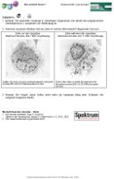
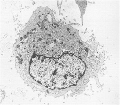
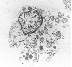
2. Welches Apoptose-Stadium hat die Zelle im rechten Bild erreicht? Begründen Sie kurz.
|
|
|
Zelle vor der Apoptose (Elektronenmikroskop. Bild: 7.000x Vergrößerung) |
Zelle während der Apoptose (Elektronenmikroskop. Bild: 11.600x Vergrößerung) |
| Quelle: http://edoc.hu-berlin.de/dissertationen/henschke-cornelia-2001-02-23/HTML/henschke-ch3.html |
Quelle:
|
3. Nennen Sie Folgen, wenn Zellen nicht mehr zur Apoptose fähig sind. Erläutern Sie mögliche Ursachen hierfür.
Weiterführende Literatur - Links:
- Zell-Harakiri auf Befehl, Peter H. Krammer, Spektrum der Wissenschaft, Spezial 3: Krebsmedizin II, S. 28-31, 2003
- http://www.biochemweb.org/apoptosis.shtm